Chanterelle Mushrooms

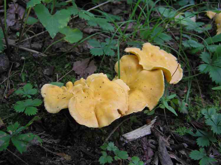
The golden-yellow hue of the chanterelle beckons the fortunate forager

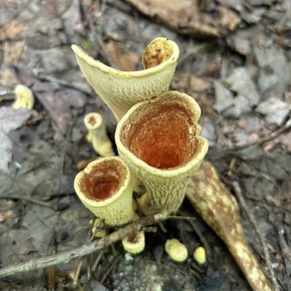
Common Name: Chanterelle, Golden chanterelle, Smooth chanterelle– Of French origin from Latin cantharis, a drinking vessel. Derived from Greek kantharos, more specific as “a deep cup of ancient Greece with a high stem … continuing the curve of the bottom of the body and rising above the rim” [1] This describes the shape of the mushroom with its cup-shaped cap and decurrent ridges that extend down the stem. Perhaps not inconsequently, chanterelle is also a French word for the highest string of a piano. The verb chanter meaning to sing, an apt metaphor for the many positive qualities of chanterelles.

Scientific Name: Cantharellus cibarius – The genus name is from cup as above. The species name is from the Latin meaning “relating to food.” It is thought to be derived from the Greek dialectal  kibba, an animal’s fodder bag, emphasizing the historical tradition of chanterelles as choice edible mushrooms. C. cibarius was once treated as a single species. Due to DNA revelations and consequent renaissance of biology, the golden chanterelle is now considered to be a complex of related but different species, following the trend of many mushrooms that must comply with the hard-wired reality of genetics. 

Potpourri: Chanterelles are likely the most well-known of edible fungi across the globe, notably in North America and Europe but almost equally in Asia and Africa. This is due to four factors. The most significant is that they are relatively easy to find. The bright yellow orange of the cap stands out against the green undergrowth of forest understory where they thrive. Of equal importance is that chanterelles are distinctive enough to allow for positive identification even among neophyte mushroom hunters. The erstwhile doppelgänger Jack-O-Lantern is only a minor impediment to confident identification, a necessity for mushroom consumption. The third factor is that they frequently fruit in extensive flushes that can blanket the ground, providing a veritable cornucopia for the gatherer. And last but not least, they are consistently delectable, choice even, and can be eaten as a side dish or added to soups, salads, or just about anything else.

The most distinctive feature of mushrooms in the Chanterelle Family Cantharellaceae is that they have ridges instead of gills, like most agarics, or tubes extending inward from the visible pores, like boletes and bracket fungi. The ridges of the chanterelles are similar in form and function to the surfaces of coral and tooth fungi in that they support the hymenium, the name for the all-important spore bearing or fertile layer of a fungus. The sole function of the mushroom, the fruiting body of the fungus, is to produce reproductive spores. Since living things must reproduce to perpetuate their genes, fungi in general evolved to produce as many spores as possible. Gills and pores are configured to maximize the surface area of the hymenium, which covers both sides of the gill or the entire circumference of the tube. A spore on a vertical gill or circumferential tube surface cannot simply be released as it would get stuck to the surface. The evolved mechanism to shoot the spore away from the hymenium involves a drop of water condensed from atmospheric moisture under the cap (which is one reason why rain is important for mushrooms). Called a surface tension catapult, the drop’s adherence to the surface of the spore propels it at a speed of about 10 miles per hour with an acceleration of some 25,000 times the force of gravity called G-Force or simply G’s (see HNB article on Parasol Mushrooms for more details). [2]
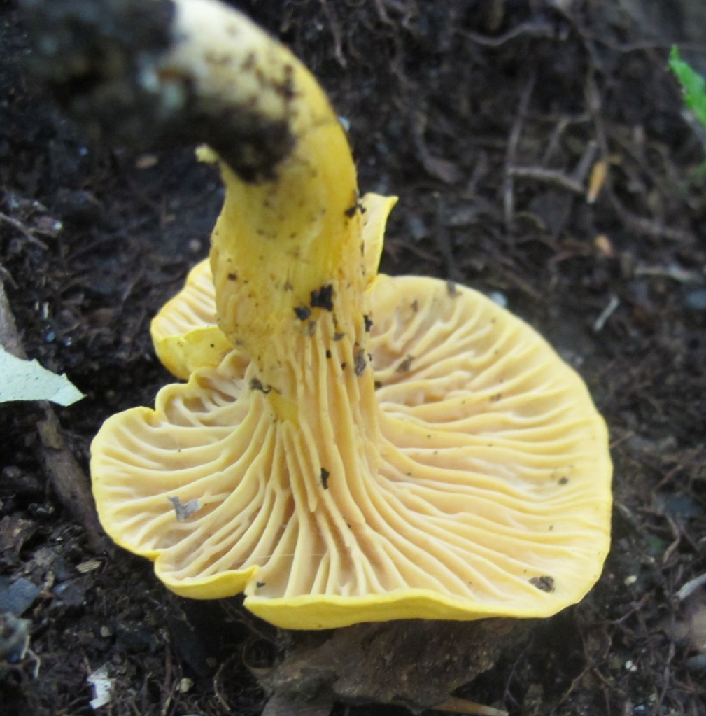
Decurrent ridges are the hallmark of chanterelles

Chanterelles and other fungi with the hymenium extending across a relatively flat (albeit ridged) surface followed a different evolutionary path. It is generally accepted that this surface spore dispersion physiology is more primitive than that of the gilled and pored fungi. This would follow the logical path of progression from producing a limited number of spores on a flat surface to the much more efficient and prolific spore production of gills and pores. The spore production dichotomy is also reflected in the relative growth and maturity of chanterelle family fruiting bodies. Whereas most mushrooms come up overnight and open almost immediately to release a torrent of spores, deliquescing to muck in about a week, chanterelles grow slowly, about one inch per month, and persist for up to three months. Chanterelles thus produce mature spores slowly over a much longer time frame and in significantly smaller numbers. [3] This raises (at least) two questions: (1) Since the fruiting bodies of chanterelles persist for so long, how do they keep from getting eaten by animals, notably insects; and (2) Since they produce a relatively limited number of spores, how can chanterelles compete successfully in an environment shared with the multitudes of spores created by all of the other fungi that specialize in spore saturation?

To answer the first question, field observation confirms that chanterelles are rarely subject to significant predation. They are almost always found intact with none of the obvious scars and torn edges that are indicative of incision by insects. In contrast, most of the other mushrooms encountered on woodland byways have pieces missing, notably the gilled mushrooms of the Russula genus and the mushrooms with pores collectively called boletes. When collected chanterelles are sorted, the older ones are excluded from the cookpot due only to general degradation in the form of dryness and chalking and not due to embedded bugs. A study conducted in Finland where chanterelles proliferate found that only one percent of chanterelles had insect larvae compared to over fifty percent for other mushrooms.  The only reasonable explanation for this observation is that chanterelles have evolved some form of chemical compound that acts as a deterrent to consumption by animals. This is hard to reconcile with the appeal of chanterelles to human animals. The delicate fruity smell of chanterelles, usually compared to apricots, is retained on cooking to impart a similar taste when eaten. [4] It is hard to imagine how an apricot aroma would deter animals, humans excepted. It is an as yet unresolved conundrum.

The second question concerns the ability of spore-sparse chanterelles to compete with spore-sporting fungi. It is related to the degree and extent of the association between trees of the forest and fungal mycelia. The mycelium is the corpus of the fungus from which the mushroom fruiting bodies sprout to sporulate. Mycelia consist of bundles of the root-like tendrils called hyphae that emerge from spores when they germinate. Since fungi are heterotrophic, they must locate and metabolize nutrients from external sources. Only plants are autotrophic. Many if not most fungi that produce mushrooms form a mutualistic relationship with trees called mycorrhizal, literally “fungus root” in Greek. Fungal hyphae surround the roots of trees in a sheath, a configuration called ectomycorrhizal. At the molecular level, the fungus provides minerals, notably phosphorous, and water to the tree in return for the hydrocarbons produced by photosynthesis. Chanterelles compete well with other forest mycorrhizal mushrooms in spite of slow growth and fewer spores. This is in part because they are masters of mycorrhizal matchups.  Tolerant of soil variance that can range from limestone to weathered granite, they form partnerships with many different tree types. The golden chanterelle, C. cibarius, has been found to associate with 14 different tree genera ranging from pines to oaks. [5] This relationship is long term − chanterelles can be found in the same areas because they have been nourished by the same forest for decades.

The global renown of chanterelles is due to their reputation among gourmands as exceptional ingredients to any cuisine. This is in no part diminished by the fact that they are not cultivated and must therefore be hand-picked by foragers who know where to find them. It has even been suggested that one way to gain the gratitude of gourmet chefs is to arrive at their restaurant with a basket of harvested chanterelles to be prepared for a meal, leaving the excess for the use of the chef in other dishes. [6] The mycorrhizal relationship between chanterelles and trees is complex. and, although there has been some modest success in promoting chanterelle growth in dedicated plantations inoculated with spawn, it has been largely experimental and not scalable. This is contrary to the common wisdom that they are “easy to transplant within congenial habitats by mycelium or spores.” [7] Recipes for chanterelles abound, ranging from sauteed in oil and seasoned with salt and pepper to Venetian “Chanterelles Risi e Bisi” (rice and peas) and Mid-Atlantic “Chanterelle Sea Food Stew.” [8]

The demand for chanterelles often exceeds the local supply resulting in national and even international trade to reduce deficits. Germany is the epicenter of both chanterelle consumption and depletion.  While most of German imports come from Lithuania, Poland. and Belarus, five million pounds, come from the Pacific Northwest of the United States. [9] This is a small fraction of the annual international trade in chanterelles, estimated at over 400 million pounds. Where trade is reduced or restricted, local demand can result in depletion, threatening local populations. For example, in Saarbrucken, Germany, seven tons of chanterelles were sold annually in the mid 1950’s. By 1970, the local harvest had plummeted to half a ton. While the decline in chanterelles is to some extent a result of overconsumption, there a general decline of mycorrhizal fungal globally due to habitat loss as forests are cleared for agriculture or urban expansion. A survey conducted in the Netherlands found that between 1972 and 1989 the average number of mushrooms in a 1000 square meter plot fell from 4720 to 370. [10] This has in part been compensated by the surge in production of cultivated mushrooms like button mushrooms and shitakes. Uncultivated chanterelles are in a category of their own in being totally dependent on nature vice nurture.
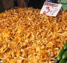
A market in Helsinki, Finland

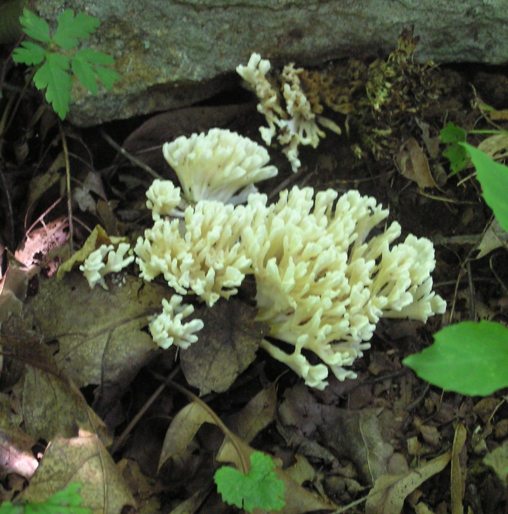
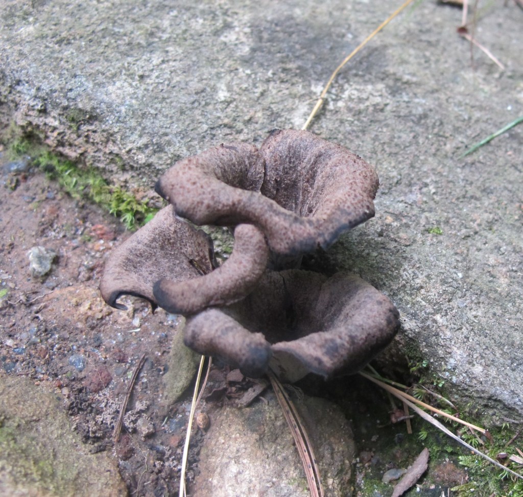
The chanterelle family Cantharellaceae is comprised of genetically related mushrooms that evolved with ridges instead of gills as the reproductive spore bearing surface. While the golden chanterelle (C. cibarius) is the most common, the Cinnabar-red Chanterelle (C. cinnararinus) is a close second and is also edible. Found in expansive tracts in open oak woods, it is rarely harvested due to its diminutive size with a cap diameter that rarely exceeds one centimeter. The Black Trumpet (Craterellus fallax) which shares the highest gastronomic encomium as “choice” like the golden chanterelle, is less common and difficult to spot due to its monochromatic blackness. However, its fruity smell is pungent, providing a second sensory vector for localization. Not all chanterelles are edible. The Scaly Vase Chanterelle (Gomphus floccosus) looks like a chanterelle that never opened, frozen in position without the namesake cup shape. It contains indigestible acids that convey a sour taste making it unpalatable. [11]
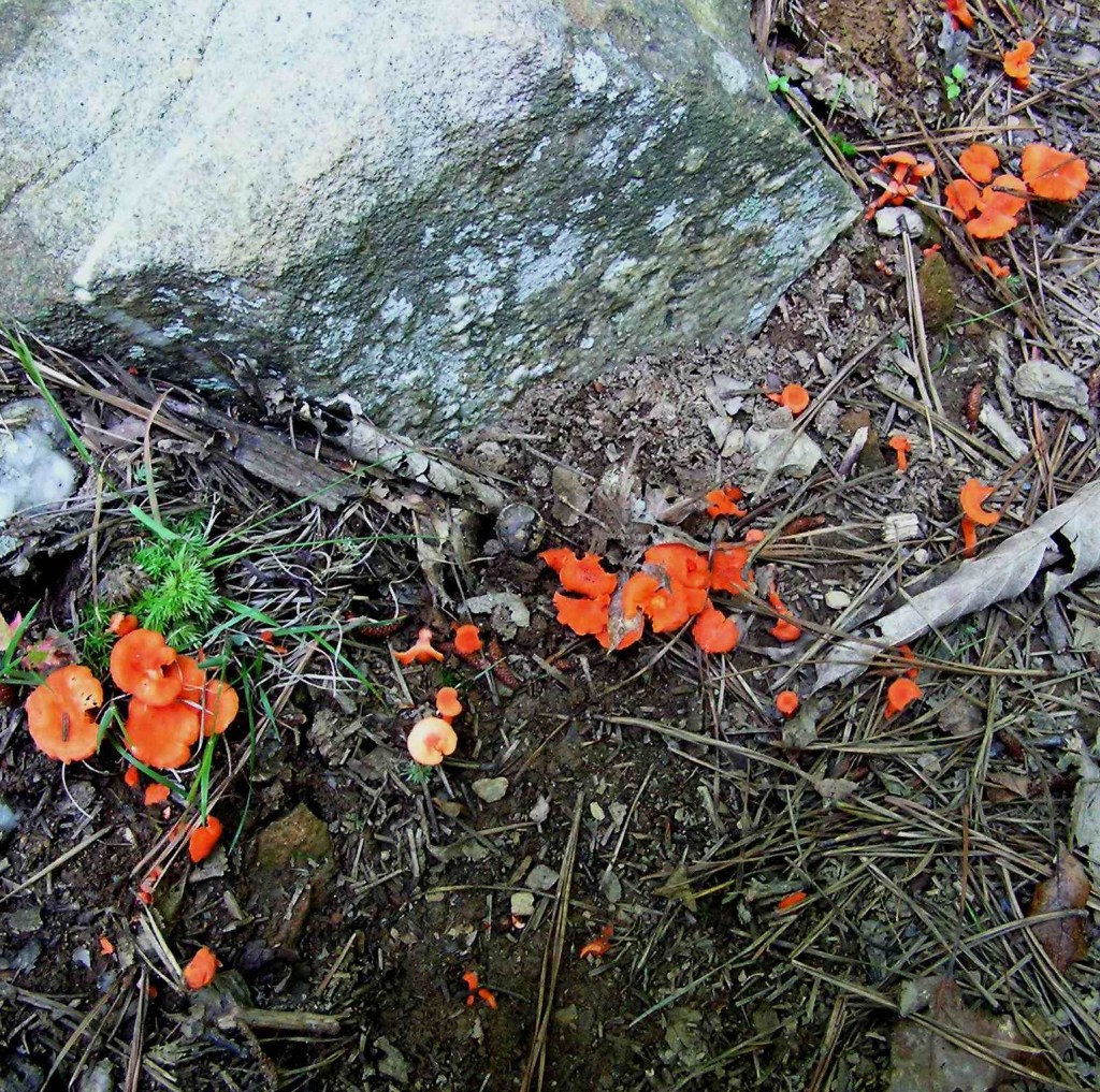
Cinnabar-red Chanterelle
Black Trumpet
Scaly Vase Chanterelle

Because of the global reach of chanterelles among diverse human populations for millennia, local traditions have led to an array of descriptive common names. Language traditions predate the establishment of modern nation states resulting in substantial variance in one area or language. In German, the names include pfifferling, pepper mushroom,  dotterpilz, egg yolk mushroom, and hasenöhrlein, little hare ear. The French don’t call it chanterelle, in spite of its Latin etymology. It has many other names instead, ranging from chevrette, small goat, and crete du coq, cock’s crest, to girolle, twisted mushroom. From the Netherlands north into Scandinavia, it is typically referred to as simply kantarell, a Germanic derivative of chanterelle. China has a long cultural history that involves fungi that probably reaches into the Neolithic Age. Chanterelles were not only consumed, as jiyou-jun, chicken fat mushroom, or xingjun, apricot mushroom, but also used to treat dry skin and prevent night blindness. [12]  Suffice it to say that chanterelles are a remarkably global species.

Chanterelles are not only nutritious but contain several important medicinal compounds. A commonly referenced quote from a 19th century European source asserts that “Not only this same fungus [the chanterelle] never did any harm (sic), but it might even restore the dead.”  [13] This is evidence of a global consensus that chanterelles and ambrosia are synonymous. Like most mushrooms, chanterelles have high levels of protein (about 10 percent by dry weight), potassium (5 percent) and nitrogen (3 percent) in addition to the fiber-rich chitin that is the structural building block of fungal cells (as cellulose is for plants). Chanterelles have levels of vitamin D comparable to the inimitable cod liver oil, and vitamin A, a result of the beta-carotenes responsible for the yellow and red hues − an antidote for vision problems like night blindness. [14] It is no small wonder then that evocative joy emanates from mushroom foragers on discovering a chanterelle expanse along a forest corridor. They may even break into song.

References:

1. Webster’s Third New International Dictionary of the English Language, Unabridged. G. C. Merriam-Webster Co. New York, 1966.

2. Kendrick, B. The Fifth Kingdom, Focus Publishing, Newburyport, Massachusetts, 2000, pp 80-98.

3. Pilz, D. et al “Ecology and Management of Commercially Harvested Chanterelle Mushrooms” USDA General Technical Report,  PNW-GTR 576. March 2003 https://research.fs.usda.gov/download/treesearch/5298.pdf   

4. Schaechter, E. In the Company of Mushrooms, Harvard University Press, Cambridge, Massachusetts, 1997, pp 132-133.

5. Pilz, D. et al op. cit.

6. Russell, B. Field Guide to Wild Mushrooms of Pennsylvania and the Mid-Atlantic, Keystone Books, Pennsylvania State University Press, University Park, Pennsylvania, 2006.pp 100-101.

7.McIlvane, C and Macadam, R. One Thousand American Fungi, Dover Publications, New York, 1973, pp 214-220.

8. Casciero, A. “A Chanterelle Extravaganza” Fungi Magazine, Volume 10 Number 2, Summer 2017. Pp 39-42

9 Arora, D. Mushrooms Demystified, 2nd Ed., Ten Speed Press, Berkely, California, 1986, p. 658.

10. Carlile, M., Watkinson, S., and Gooday, G. The Fungi, Elsevier Academic Press, London, 2001, pp354-355.

11. Lincoff, G. National Audubon Society  Field Guide to North American Mushrooms, Alfred A. Knopf, New York, 2002, pp 387-398.

12. Pilz, D. et al op. cit.

13. Trattinick, L. 1764-1849  Die essbaren schwmme des Oesterreichischen Kaiserstaates (which translates literally as Edible Sponges of the Austrian States – probably dialect for mushrooms) https://archive.org/details/dieessbarenschwm00trat/page/n35/mode/2up  (often quoted in numerous sources)

14. Pilz, D. er al op. cit.
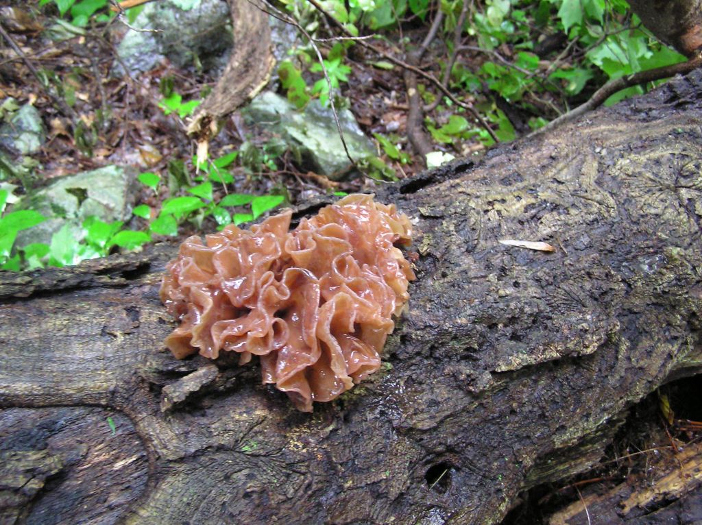
Jelly Fungi

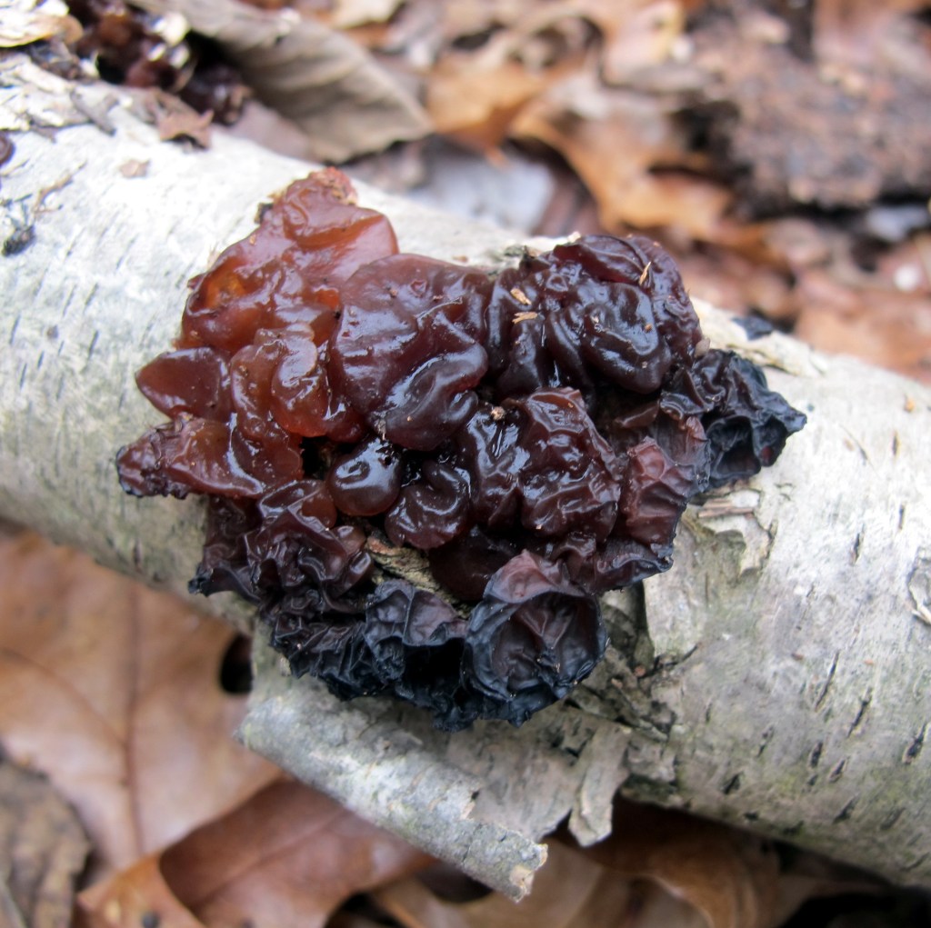
Jelly Fungus is one of the more appropriate common names

Common Name: Jelly Fungus – The gelatinous texture of the fruiting body gives rise to the general jelly description which implies being soft but resiliently firm, partially transparent, and semisolid. While the jelly appearance is true of many of the species, it is a widely inclusive designation. Brain-like, scattered blobs, and shaped like a leaf or an ear are descriptions that would better describe individual species. The species above is commonly called Jelly Leaf, or, alternatively, Brown Witch’s Butter as an example of confusing fungal terminology.

Scientific Name:  Tremellales, Dacrymycetales, Auriculariales and Sebacinales are the four Orders into which jelly fungi have been taxonomically assigned for now. The Latin word for tremble is tremulus giving rise the trembling order. Dacry is Latin for tears, implying wetness. Auricula is Latin for ear, here used for the shape of the fungus. Sebaceus is Latin for tallow which would imply a fatty appearance. The jelly fungus above is Tremella foliacea, which translates from Latin to something that approximates “tremulous leaf.”

PotpourriJelly fungi, while of marginal importance to the ecological network of the forest,   attract the inordinate attention of hikers passing through. They come in a variety of colors, striking many as an unlikely yet obvious protuberance on fallen logs. The key attraction is the gelatinous texture of the overlapping random lobes and curls, suggestive of an underlying reason for their topology.  And there is one. Like all fungi, they are the fruiting bodies of the fungal mycelium that is embedded in the rotted wood (and in some cases on the backs of other, parasitized, fungi) on which they appear. The only function of the jellied fruiting bodies is to provide an effective means to launch as many of their reproductive spores into the air to propagate the species.  

The taxonomy or scientific classification of those species of fungi that are jelly-like in appearance has not yet been settled conclusively. The DNA research necessary to establish phylogenic assignations has understandably been sparse due to both the obscurity and relative insignificance of lumps of colored jelly.  For this reason, they are almost always combined in field guides as a single category entitled jelly fungi since they are visually distinctive.[1] However, there are some details that are germane. Jelly fungi are all in the largest class of fungi that includes the well-known gilled mushrooms named Basidiomycota, named for the basidia, the structures on which the reproductive spores are produced. Jelly fungi have at least one  physiological features that distinguish them within the larger class. Along with rusts and smuts, they have basidia that are divided into sections by a cellular wall. They are therefore  called septate or divided basidia basidiomycetes and are grouped in the subclass Heterobasidiomycetes (this was at one point established as a class named Phragmobasidiomycota along the endless timeline of sorting out fungal genetics). [2] The idea is that they are different (hetero) from the rest of the class that are, not surprisingly, called Homobasidiomycetes.

While the esoterica of fungal taxonomy establishes the credentials of biological determinism, it hardly satisfies the practitioner. After all, Carl Linnaeus created “the road map of biology” four centuries ago based entirely on physical appearance. The fungal details were expanded some years later by another Swede named Elias Magnus Fries and acclaimed as “the Linnaeus of Fungi”.  Reproduction is what creates and perpetuates living things so taxonomy focuses on the details of its execution. For fungi, it is all about how and where the spores are created. The evolution of fungi over time or phylogeny is then at least in part about better ways to form and release spores into the environment. From the phylogenic perspective, jelly fungi constitute an early experiment with spore dispersal that gave way over time to the evolution of mushroom shaped structures with gills and pores as improvements. The mushroom shape of many fungi that succeeded the jellies improves on the crucial propagative function with basidia extending outward from the sides of gills or pores for better dispersal. While the jellies survived, mushrooms thrived. [3]

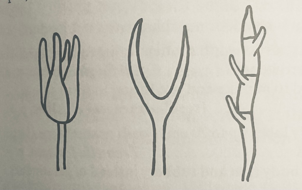
While speculative, it is plausible that the jelly-like structure evolved in an area or region with intermittent and restricted rainfall, as it desiccates when dry and revives on the restoration of moisture. This would provide for the release of spores only after a rain to better promote spore dispersal and germination. Most if not all fungi have a method to control the release of spores to coincide with the favorable environmental conditions of moisture. The different shapes of the basidia in the various orders of jelly fungi all serve to extend the spore formation and dispersal structure, the basidia, through the jelly to reach open air for wind dispersal. It is probable that the different shapes of the basidia were due to the classic Darwinian process of random mutation producing variants that on some relatively rare occasions, led to an improvement in the overall population of the new genetic code map. The longitudinally septate basidium on the left defines the order Tremellales, the forked shape in the middle is Dacrymycetales, and the transversely septate arrangement is Auriculariales. [4]

Septate basidia of three orders of jelly fungi

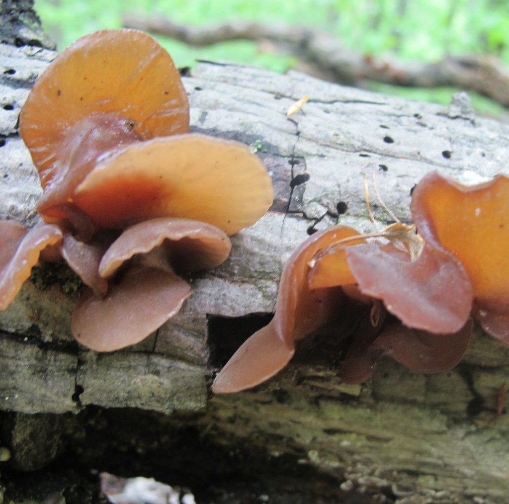
The edibility of jelly fungi has long been a matter of conjecture. While jelly implies edible and no jelly fungi are known to be poisonous, there is scant evidence one way or the other. This is certainly in part due to their slimy texture that would more likely repel than attract the fungivore. One field guide distinguishes those jelly fungi listed as either inedible or nonpoisonous. [5] It is not clear what criterion might have been used to establish the distinction. There is one jelly fungus that is universally recognized as edible. Auricularia auricula (literally ear-ear in Latin) commonly called  wood ear, tree ear, or Judas’ ear, is common globally across the northern hemisphere. It is the earliest known fungus to have been grown commercially, dating from 600 AD in China where it is known as Mu Ehr. In 1997, it was the fourth most important edible mushroom in the world. [6] It is still at the forefront of cultivated edible fungi.

Tree Ear (Auricularia auricula)

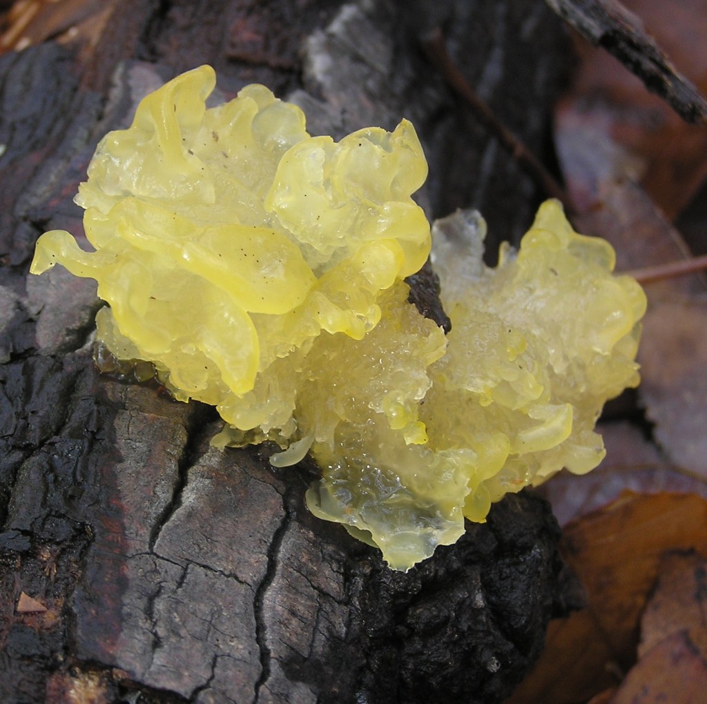
Witch’s Butter (Tremella mesenterica) is second only to tree ear among known and recognized jelly fungi. Both the common name and the scientific name exemplify the fanciful names assigned by both custom and intention. Since it is yellow, the butter aspect is unremarkable, but since it is found randomly growing in dense woods, ascribing it to the dark and usually evil connivances of witches is metaphorical. The species name mesenterica  translates as middle intestine, which, while descriptive, connotes for most people an aversion to any consideration of edibility. The supernatural association is enhanced by the physical nature of the fungus, which will desiccate to become indistinguishable only to come coursing back to full prominence when moistened, or perhaps by some incantation. [7]

Witch’s Butter (Tremella mesenterica)

Dead Man’s Rubber Glove (Tremella reticulata) is another of the jelly fungi with a common name that overemphasizes a descriptive mnemonic at the expense of any consideration as an edible. It is also called White Coral Jelly due to its similarity in basic structure to the coral fungi characterized by upright, branched lobes. In both cases (jelly and coral), branching and widening produces more surface area over which to distribute the spore forming hymenium. As an example of the nomenclature variability among jelly fungi, Jellied False Coral, almost identical in appearance to the others, is listed in a more recent field guide as Tremellodendron schweinitzii. This same reference lists T. reticulata as Sebacina sparassoidea in pointing out that species of the Tremella genus are parasitic on other fungi, mostly those of the genus Stereum, whereas T. schweinitzii is mycorrhizal. [8] The study of jelly fungi continues as details of its taxonomy are revealed.[9]

Dead Man’s Rubber Glove (Tremella reticulata)

The most stygian of all the jelly fungi is Exidia glandulosa, commonly called Black Jelly Roll. Not surprisingly they are also called Black Witch’s Butter, or, in the UK, simply Witch’s Butter, with the presumption that any butter from witches would be black. E. glandulosa is closely related to the edible tree ear which raises the inevitable edibility question among those who fancy fungi (second only to the “is it poisonous?” question). Those field guides that include it are either  silent on the subject (assuming no one would want to know), or pointedly facetious in noting that its edibility is “unknown, and like most of us, likely to remain so”. [10]

Black Jelly Role or Black Witch’s Butter (Exidia glandulosa)

Or perhaps we should give jelly fungi another look as a culinary choice. Providing nutritious food to the burgeoning human population is one of the challenges of the current century.  Tree ear (A. auricula) has been systematically analyzed as the only jelly fungus that is currently cultivated and consumed and found to be highly nutritious. One hundred grams of dried tree ear consists of over four grams of protein and almost twenty grams of fiber with 351 calories of energy. [11] It has been suggested, albeit by a decidedly outré American mycologist, that “old tradition, in many countries, attests that the Tremellas are Fairy bread” and that “pretty indeed must have been the feasts when piles of such purity filled the board.” Noting that some jelly fungi are “delicious when slowly stewed,” and that others are  “gelatinous, tender – like calf’s head,” it is held that all fungi, save the poisonous Amanitas, deserve a taste. [12] There is a lot to learn about the Kingdom Fungi, as it takes its rightful biological place beside plants and animals, maybe even food for thought.

References:

1. Lincoff, G. National Audubon Society Field Guide to North American Mushrooms, Alfred A. Knopf, New York, 1981. pp 379-386.

2. Kendrick, B. The Fifth Kingdom, Third Edition, Focus Publishing, R. Pullins Company, Newburyport, Massachusetts, 2000, pp 82-83, 102-103.

3. Stevenson, S. The Kingdom Fungi, Timber Press, Portland, Oregon, 2010. pp 100-127.

4. Arora D. Mushrooms Demystified, Second Edition, Ten Speed Press, Berkeley, California, 1986. pp 669-675.

5. Miller, O. and Miller H. North American Mushrooms, A Field Guide to Edible and Inedible Fungi, Morris Book Publishing, Guilford, Connecticut, 2010, pp 491-498.

6. Chang, S. and Miles, P. Mushrooms, Cultivation, Nutritional Value, Medicinal Effect, and Environmental Impact, Second Edition, CRC Press, New York, 2004, p 206.

7. Roody, W. Mushrooms of West Virginia and the Central Appalachians, The University Press of Kentucky, Lexington, Kentucky, 2003, pp. 452-455.

8. Baroni, T. Mushrooms of the Northeastern United States and Eastern Canada, Timber Press, Portland Oregon, 2017 pp 510-519.

9. Hyde, K.  et al. “Towards an integrated phylogenetic classification of the Tremellomycetes”  Mycosphere. 8 January 2016  Volume 15 Number 1 pp  5146–6239.

10 Arora D. op cit. p 673.

11. Chang, S. and Miles, P. op cit. p 29.

12. McIlvane, C. and Macadam, R. One Thousand American Fungi, General Publishing Company, Toronto, Canada, 1973, pp 526-531

Destroying Angel – Amanita bisporigera

The key features of the Destroying Angel are the cup-like volva at the base of the stem, the stark whiteness of the stem, cap, and gills, and the partial veil hanging from the top of the stem just below the gills under the cap.

Common Name: Destroying Angel, Fool’s Mushroom, Death Angel, White Death Cap – The virginal whiteness of all parts of the mushroom are aptly described as angelic – beautiful, good, and innocent. The fact that it is anything but is conveyed by the addition of destroying with death-dealing toxicity.

Scientific Name: Amanita bisporigera – The generic name is taken directly from the Greek word amanitai, probably from the Amanus Mountains of southern Turkey where the noted Greek physician Galen may first have been identified the archetype, Amanita. [1] The specific name indicates that there are only two spores on each of its basidia in contrast to the four spores of other basidiomycete fungi. Virtually indistinguishable from Amanita virosa and Amanita verna which both frequently appear as synonyms in mushroom field guides.

Potpourri:  The destroying angel is a toadstool nonpareil. While the origin of the term toadstool is obscure, it cannot be a coincidence that tode stuhl means death chair in German, the language of the Saxons who emigrated to England. Its notoriety is not only because it is one of several mushrooms that contain deadly poisons called amatoxins, but also due to its close resemblance to Agaricus campestris, the edible field mushroom which is the cousin of the cultivated white button mushroom of supermarkets and salad bars. Both are white, similar in size and shape, and grow in the same habitat, primarily grass under or near trees. The destroying angel is the most dangerous of the numerous doppelgänger mushrooms as the deadly twin of a well-known and often consumed edible.  Misidentification absent knowledge of the subtle physical differences between the two can result in discovering the profound physiological differences with sometimes deadly result. The field white mushroom is nourishing. The angelic white mushroom is Shiva.

The cup at the bottom of the stem is the volva, the bottom half of the universal veil.

The key features that distinguish the destroying angel from similar mushrooms are straightforward if you know what to look for. First and foremost is the volva, (Latin for a covering like a husk or shell) which is the cuplike structure at the base of and surrounding the stem or stipe. The volva is frequently hypogeal, i. e. underground and out of sight. This means that it can only be positively identified by digging up the soil around the base of the mushroom. [2] However, it is the standard and preferred practice among mushroom gatherers to use a knife to cut through the stem cleanly at the base. This is done so the mycelium of the fungus from which the fruiting body mushroom grows is not seriously disturbed. The procedure is analogous to gathering apples from an apple tree. The fungal mycelium and the apple tree survive to produce new mushroom spores and fruit seeds for future generations. Using the standard harvesting technique, it is easy to see how the below the cut volva would not be noted.  White mushrooms must be dug out to the roots to avoid the dilemma of the death mushroom.

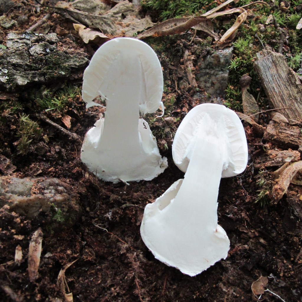
The only way to be certain that you have a puffball and not a Destroying Angel is to cut it in half.

The volva is the bottom part of what is known as a universal veil, a thin membrane that envelops the mushroom during the subterranean growth phase to protect the gills and the spores they hold from damage. The universal veil is a characteristic of all mushrooms in the Amanita Family. While there are a few other mushrooms that have a universal veil and its volva (such as the genus Volvariella named for this characteristic feature), it is a reliable identification feature for the destroying angel. All spore-bearing mushrooms are produced by the fungal mycelium underground as an ovoid called a primordium. Once they mature and environmental conditions are promising (like after rain) the extension of the stem causes the universal veil to tear around its circumference to expose the cap and gills of the fruiting body for spore dispersal. The volva is the lower part of the “eggshell” that remains attached to the bottom of the stem. Prior to upward extension, the destroying angel looks like a white egg, similar in appearance to a puffball, another type of edible fungus with which the destroying angel can be confused.  Some field guides include a picture of it in the puffball section to emphasize the danger of mistaken identity. [3] The only way to be absolutely sure is to cut the fungus lengthwise to reveal a cap and gills within.

Many mushrooms have what is known as a partial veil which also helps prevent damage to the reproductive gill surface. It is partial in that it only covers the underside of the cap, extending from the edges of the cap to the stem. When the mushroom cap expands fully, the partial veil also tears, in many cases leaving some remnants around the edges and a ring called an annulus attached to the stem just below the cap. In some cases, the partial veil remnant can be seen hanging like a draped clerical mozetta at the top of the stem. However, this annular ring is not well connected, and in many mushrooms with partial veils, there is no remnant. Most Amanita family mushrooms have both universal veils and partial veils with both a volva at the bottom and a ring around the stem as is the case with the destroying angel. The double protection afforded to the gills must have evolved due to the success of the species in propagation. Amanitas are one of the most prolific of all mushroom families. Partial veils and the remnant annulus are also a characteristic of the Agaricus family, which includes the edible field mushroom Agaricus campestris. They do not have universal veils with the tell tale volva.

The second prominent feature of the destroying angel is the stark whiteness of the cap, stem, and gills that has been described as having a “strange luminous aura that draws the eye” that is “easily visible from one hundred feet away with its serene, sinister, angelic radiance.” [4] The cap is smooth and usually described as viscid or tacky when wet.  This is to distinguish it from most of the other species in the Amanita genus that have warty patches on the cap from the dried out and cracking universal veil like the white dot warts on the bright red cap of the iconic fly agaric (Amanita muscaria).  The glowing purity of the whiteness is a reliable feature for initial field identification. Confirmation by looking for a picture or drawing of a white mushroom with a volva and annular stem ring using a field guide is another matter. One provides only Amanita verna or fool’s mushroom, prevalent only in spring (vernus in Latin). The common name implies that it fools the observer with its deception. [5] A second field guide provides both A. verna as the spring destroying angel, and Amanita virosa (virosus is poisonous in Latin) for mushrooms that appear in the fall with only a passing reference to A. bisporigera. [6] DNA sequencing of fungi has had a profound impact on the eighteenth-century Linnaean system basing taxonomy on physical similarity. It has been shown that all destroying angels of North America are A. bisporigera (with one additional species A. ocreata in California) and that A. verna and A. virosa are only found in Eurasia. Destroying angel is a universal common name for all species for the white mushrooms with volva.

The destroying angel is one of the deadliest mushrooms known. According to one account “misused as a cooking ingredient, its alabaster flesh has wiped out whole families.” [7] The toxic chemicals are called amatoxins (from the generic name Amanita), which are protein molecules made up of eight amino acids in a ring called a cyclopeptide with a molecular weight of about 900. The death dealing amatoxin variant is alpha-amanitin, which destroys RNA polymerase, a crucial metabolic enzyme. RNA polymerase transcribes the DNA blueprint, creating  messenger RNA that transport the codon amino acid recipe used  to make proteins on which all life depends. The ultimate result is rapid cell death. The gastrointestinal mucosa cells of the stomach, the hepatocytes of the liver, and the renal tubular cells of the kidneys are the most severely affected cells because they have the highest turnover rate and are rapidly depleted. The liver is most at risk because the hepatocytes that absorb alpha-amanitins are excreted with the bile and then reabsorbed. The initial stages of amatoxin poisoning start about ten hours after ingestion; the gastrointestinal mucosa cells are the first to be affected resulting in forcible eviction (aka vomiting) of the intruding poisons.  There follows a period of several days of calm as the stomach cells recover somewhat before the storm of  hepatic and renal debilitation. The third and final stage can in severe cases lead to the crescendo of convulsions, coma and death. The lethal dose for 50 percent of the population or LD50 is used by toxicologists as a benchmark for relative virulence. The LD50 for alpha-amanitin is 0.1 mg/kg.  A 70 kg adult will have a 50-50 chance of survival with a dose of 7 milligrams, the amount of alpha-amanitin in one small destroying angel. [8]

The North American Mycological Association (NAMA) received a total of 126 reports of amatoxin poisoning over a period of thirty years, about four annually. The fatality rate has historically been on the order of thirty percent attributed to liver and/or kidney failure; this number has improved over the last several decades to about five percent due to a better understanding of amatoxin physiology effects combined with aggressive therapy. The basic tenet of the treatment is to reduce the toxic concentration in the blood serum as rapidly as possible. Gastric lavation is used if the ingestion was recent enough followed by a thorough purging using emetics to induce vomiting and cathartics to induce evacuation of the bowels (essentially the same effect on the gastrointestinal mucosa cells to expel the poison).  Perhaps the most important therapy is the use of activated charcoal, as amatoxins have a high affinity for adsorption on its surface. Although there is no proven antidote, intravenous injections of penicillin have been used with some apparent benefit. A French physician named Bastien developed a three part procedure using intravenous injections of vitamin C and two types of  antibacterial drugs supplemented with penicillin to successfully treat 15 cases. To unequivocally prove its efficacy, he conducted the ultimate experiment by eating 70 grams of Amanita phalloides, the death cap cousin of the destroying angel and using the protocol on himself. [9] The most promising new treatment is silibinin, an extract of the blessed milk thistle (Silybum marianum), which is sold commercially as Legalon by a German pharmaceutical company. Liver transplant was once considered the last resort for amatoxin poisoning, but that may no longer be necessary. [10]

The destroying angel is not the only mushroom that produces amatoxin, nor is amatoxin the only substance produced by fungi that is inimical to humans. The identification of fungal toxins and the characterization of their imputed symptoms are among the most empirical of forensic science. The facts are based almost entirely on the anecdote. The identification of the mushroom that caused the condition under evaluation is usually a matter of conjecture since the victim has eaten the evidence. To add to the confusion, the alleged offending mushroom may have been consumed with a mixture of other wild foods and fungi gathered over a wide area in obscure nooks.  The dearth of fungal knowledge in the medical community contributes to uncertainly. Poison Control Centers (PCC) were established after World War II to deal with the proliferation of chemicals as clearing houses for information about poisons and their antidotes and treatment protocols. [11] Over the ensuing years, mushroom poisonings accounted for only one half of one percent of all PCC reports (1 in 200). Of those reported, only 10 percent included any information about the mushroom. Based on limited data, NAMA established a toxicology committee in 1985 and began to supplement the PCC data with a separate data base using the input from experienced mycologists and mushroom aficionados. The result to date is a more comprehensive accounting with fairly reliable identification of 80 percent of the mushrooms involved in poisoning. [12] This is a good start but has done little to assuage the beliefs of the general public that most if not all mushrooms are toadstools and that eating wild mushrooms is a fool’s errand, sometimes literally.

One example suffices to point out the irrational fear of amanita mushroom poisoning and the broader category of mycophobia. In 1991, the venerable French reference Petit Larousse Encyclopédie was recalled because the deadly amanita article lacked the appropriate symbol for poison. But this was not enough, since almost 200,000 copies had already been sold.  Several hundred students were hired to visit 6,000 stores throughout Europe and Canada to affix stickers with the appropriate symbol for poison on the pages and append a notice on the cover of the book that it was a new edition. [13]  History has impugned the mushroom as the source of the poison that has dispatched any number of notables, among them Claudius, the fourth Roman Emperor. The perpetrator is alleged to have been his fourth wife Agrippina who wanted her son Nero to succeed to the throne. The death is recounted by the philosopher Seneca the Younger in December 54 CE, only two months after the event occurred. According to his account, it happened quite quickly, the onset of illness and death being separated only by about an hour. [14] The mushroom assassination of Claudius is almost certainly apocryphal, as deadly mushrooms are relatively slow to act; those that act rapidly generally cause gastrointestinal distress that is rarely fatal. Hyperbole is not out of the question. One recent account attributes the disappearance of the Lost Colony of Roanoke to the relocation of the starving colonists to the island of Croatoan. Gorging themselves on the mushroom bounty that they found there, they died a horrible death of grotesque contortions. [15]

References:

1. McIlvaine, C. One Thousand American Fungi, Dover Publications, New York, 1973 pp 2-5

2. Roody. W. Mushrooms of West Virginia and the Central Appalachians, The University Press of Kentucky, Lexington, Kentucky, 2003, pp 62-63.

3. Lincoff, G. National Audubon Society Field Guide to North American Mushrooms, Alfred A. Knopf, New York, 1981. pp 551-552.

4. Russel, B. Field Guide to Wild Mushrooms of Pennsylvania and the Mid-Atlantic, The Penn State University Press, University Park, Pennsylvania, 1935, pp 67-69.

5. McKnight, K and McKnight, V.  Peterson Field Guide to Mushrooms of North America, Houghton Mifflin Company, Boston, 1987, pp 238-239, Plate 27.

6. Pacioni, G. (Lincoff, G, US editor) Guide to Mushrooms, Simon and Schuster, New York, 1981, pp 76-77.

7. Money, N. Mr. Bloomfield’s Orchard, Oxford University Press, Oxford. 2002 p 151

8. Hallen, H. et al. “Gene family encoding the major toxins of lethal Amanita mushrooms”. Proceedings of the National Academy of Sciences. 27 November 2007 Volume  104  Number 48  pp 19097–19101

9. Kendrick, B. The Fifth Kingdom, Focus Publishing, Newburyport, Massachusetts, 2000, pp 319-321.

10. Beug, M. in Fungi Magazine Volume 1 Number 2 Spring 2008. Beug is a Professor Emeritus at Evergreen State College and a member of the NAMA toxicology committee.

11. Wyckoff, A. “AAP Had First Hand in Poison Control Center” AAP News Sept. 2013 http://www.aappublications.org/content/34/10/45

12. Beug, M, et al “Thirty-Plus Years of Mushroom Poisoning: Summary of the Approximately 2,000 Reports in the NAMA Case Registry” Mcllvanea Volume 16 number 2 Fall 2006 pp 47-68.

13, Schaechter, E. In the Company of Mushrooms,  Harvard University Press, Cambridge, Massachusetts, 1997, pp 210-211.

14. Marmon, V. and Wiedemann, T. “The Death of Claudius” Journal of the Royal Society of Medicine, Volume 95, May 2002 pp. 260-261.

15. Spenser, S. “The First Case of Mass Mushroom Poisoning in the New World” Fungi Magazine, Volume 11, Number 4, Fall 2018, pp 30-33.